


FLIM
fluorescence lifetime imaging microscopy, TCSPC, time-correlated single photon counting, time-to-digital converter, TDC, time tagger, analog-to-digital converter, ADC, FRET, Förster resonance energy transfer, photoluminescence decay measurements
Fluorescence Lifetime Imaging Microscopy (FLIM) is an advanced optical microscopy technique that maps the spatial distribution of excited-state lifetimes (decay time τ) of fluorescent molecules rather than relying solely on fluorescence intensity.
Fluorescence lifetimes typically range from hundreds of picoseconds to several nanoseconds. Because the lifetime is fundamentally independent of fluorophore concentration, excitation intensity fluctuations, and moderate photobleaching, FLIM provides robust quantitative insight into the local cellular microenvironment (e.g., pH, viscosity, refractive index, oxygen concentration) and enables molecular interaction studies via FLIM-FRET.
Measurement Principle and Fast Data Acquisition: In scanning FLIM systems (such as confocal laser scanning or multiphoton microscopy), the sample is scanned point-by-point using a high-repetition-rate pulsed laser (typically 20 to 80 MHz). Emitted photons are detected by sensitive single-photon detectors (SPADs, PMTs, MCPs) and recorded via Time-Correlated Single Photon Counting (TCSPC):
- Picosecond Time-Tagging with TDCs: High-resolution Time-to-Digital Converters (TDCs such as xTDC4 or TimeTagger) register the precise arrival time of each photon relative to the laser sync trigger with picosecond accuracy. Auxiliary digital input channels record pixel, line, and frame markers from galvo or resonant scanners to seamlessly assign photon timestamps to spatial image coordinates.
- Digitizer-Based TCSPC with Ultra-Fast ADCs: Ultra-fast analog-to-digital converters directly sample the detector signal. By capturing the complete pulse waveform, constant-fraction discrimination (CFD) to eliminate time walk, pulse height analysis, and reliable detection of multiple events (pile-up) can be performed digitally in the FPGA. This enables the highest count rates with excellent temporal resolution.
The accumulated timestamps form a decay histogram for each pixel. Fitting algorithms extract the characteristic lifetime τ to render a color-coded 2D or 3D FLIM image.
Please also refer to our detailed description of this application.
Glossary Title Tag:
What is FLIM? (Fluorescence Lifetime Imaging Microscopy) | cronologic Glossary
Meta Description:
Fluorescence Lifetime Imaging Microscopy (FLIM) maps fluorophore decay times per pixel. Discover how high-speed TDCs and ADCs enable picosecond TCSPC imaging.
Read more about FLIM in this detailed article.
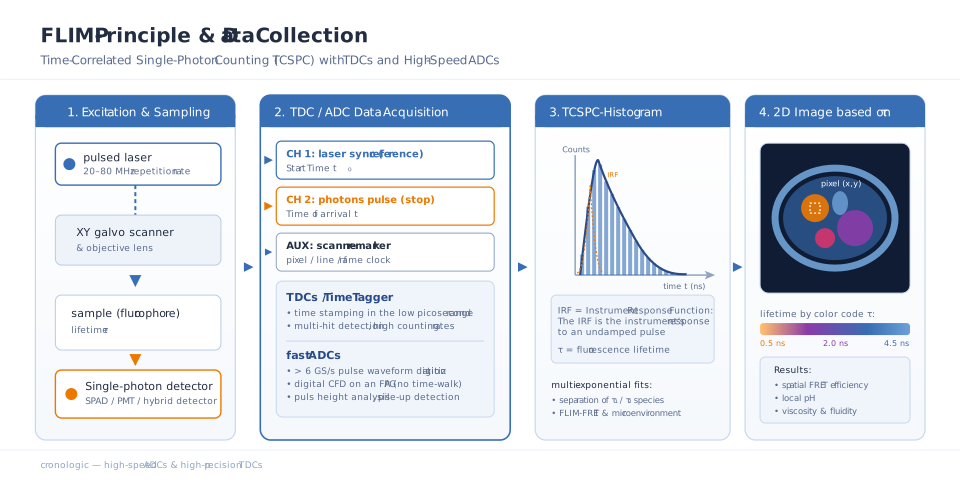
Workflow of a FLIM measurement: Time-correlated single photon counting (TCSPC) per pixel utilizing fast TDC/ADC electronics to generate spatial lifetime maps.